Innovation through academic research programs
Innovation through academic research programs
SFITS Research
The right platform for academic research programs
We support you with
- An adapted environment to test and validate your concepts
- A complete ecosystem to foster discussion, receive feedback, and further develop your projects
- A network to connect with medical experts and field specialists
- An adapted setting to give you credibility and visibility
- An adequate platform to create synergies with interventional room professionals
- A partner to participate in innovative projects: Innosuisse, Swiss National Science Foundation
The right setting for academic research activities
- Customized test environment
- Close to real-life conditions
- Safety and security
- At no patient risk
- Confidentiality
- Access to network, for direct feedback
- Experienced and professional team with specific know-how
- Ethically managed anatomical specimens
- Adapted surgical infrastructure
- High-end biomedical equipment
- Recording and video editing capacities
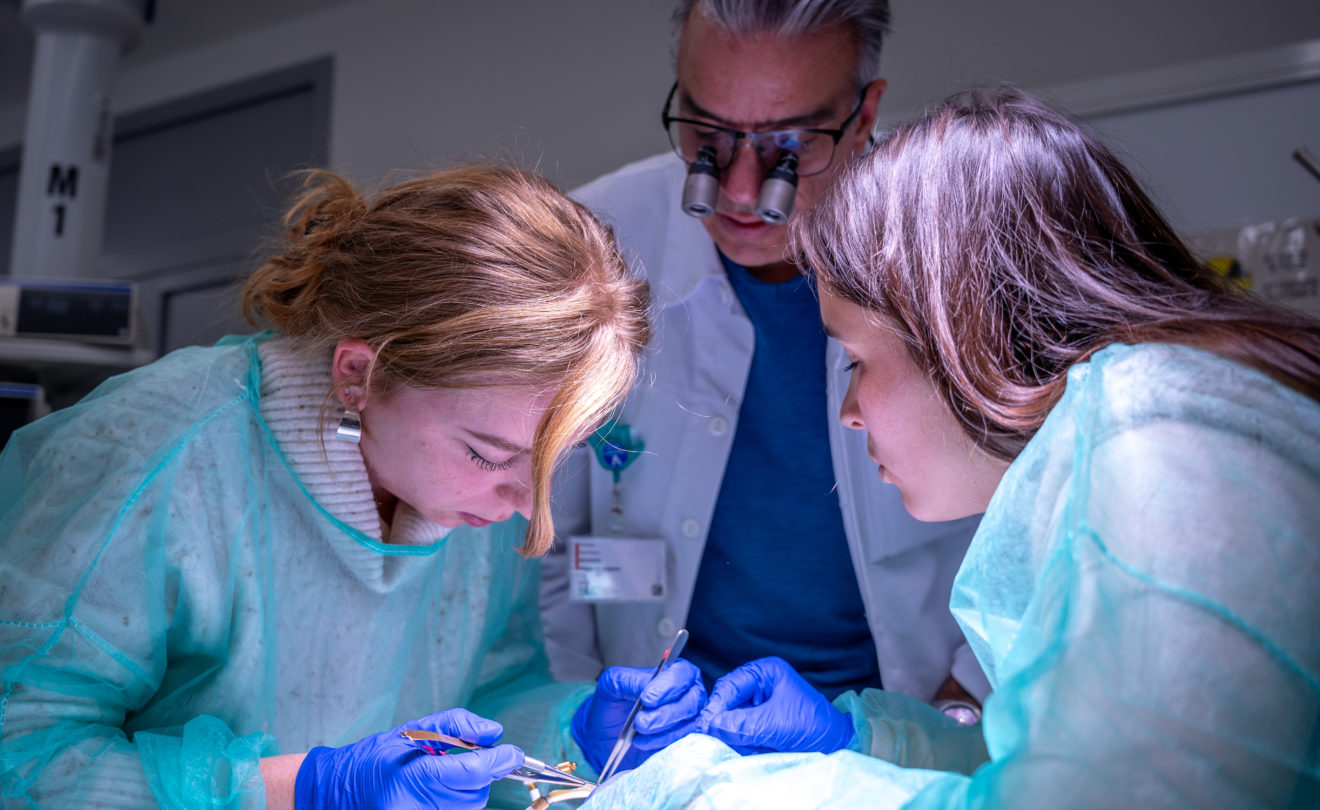
Projects and Publications carried out at the SFITS
Advanced Training on Perfused Specimen for Cardiovascular Surgeons: A new paradigm in surgery teaching
“Chargé d’enseignement et responsable de l’unité de chirurgie vasculaire au sein du service de chirurgie cardiovasculaire, j’ai développé en sus de mon activité opératoire, un réel intérêt pour l’enseignement, convaincu que la chirurgie reste fondée sur l’apprentissage par l’exemple, à la manière d’un compagnonnage.” PD Dr Nicolas Murith
Force est de constater que les outils classiques d’enseignement ont laissé leur place aux nouvelles technologies dont l’entrainement sur simulateurs. Cependant, l’apprentissage pratique reste une étape essentielle pour former des chirurgiens compétents et performants. C’est pour ça qu’aujourd’hui les exigences éthiques et le respect du patient imposent de repenser les méthodes traditionnelles d’enseignement chirurgical afin d’offrir aux jeunes médecins un entraînement à la fois réaliste, sécurisé et innovant.
Le PD Dr N. Murith décide de développer, à côté de son activité opératoire, un programme avancé de formation autour d’un concept immersif : des cours sur corps perfusé permettant de reproduire de manière fidèle les interventions courantes de la discipline.
Surgical Training for Bowel Endometriosis: A New Wet-lab Model
Dr Stas Shabanov, Dr Jean-Marie Wenger, Dr Jean Dubuisson, Prof. Patrick Dällenback, Dr Nicolas Buchs and Dr Nicola Pluchino elaborated a new training model for the management of severe cases of endometriosis with digestive involvement in particular, with the support of the SFITS.The study objective is « to demonstrate a new wet-lab model for training in conservative bowel endometriosis surgery (shaving and discoid resection) » and is now published in the prestigious Journal of Minimally Invasive Gynecology (JMIG).
This new and highly realistic model allows the next generation of endometriosis surgeons to acquire adequate training to make bowel surgery safer and more effective.
Development of an aneurysm model on human placenta for hybrid endovascular and neurosurgical training
A research project on the development of an innovative aneurysm model on human placenta for hybrid endovascular and neurosurgical training, section « education and innovation ». Some of the tests for this study were conducted at the SFITS. Led by Master Student François Kostadinov, under the supervision of Dr. Julien Haemmerli, Dr. Andrea Rosi and Prof. Philippe Bijlenga.
Intracranial aneurysms are serious vascular conditions that are difficult to treat and require highly specialized skills in both neurosurgery and interventional neuroradiology. Training for these procedures remains challenging, as current models are often constrained by ethical issues, high costs, or limited anatomical realism. In this context, the present study aims to assess whether human placenta tissue can be used to create a realistic ex vivo model of saccular aneurysms. To do so, freshly obtained placentas were circulated with a saline solution containing heparin, exposed to elastase applied locally, and then subjected to controlled internal pressure and suction in order to reproduce the mechanical conditions that promote aneurysm formation.
EEG Spatiotemporal Patterns Underlying Self-other Voice Discrimination
Prof. Karl Schaller, Giannina Rita Iannotti, Pavo Orepic and many more studied the Spatiotemporal Patterns Underlying Self-other Voice Discrimination. During their research, they combined psychophysics, voice-morphing technology, and high-density EEG to identify the pattern.
With 26 healthy participants, both with air-and bone-conducted stimuli, they found a self-voice-specific EEG topographic map occurring around 345 ms post-stimulus and activating a network involving insula, cingulate cortex, and medial temporal lobe structures. Specifically, the better participants performed at SOVD task, the less frequently they activated this network.
Neuroprosthetics research project
The “Cognitive Surgical” clinical research project will bring neurosurgery, cognitive neurosciences and neurotechnology together for the first time.
This project will aim at stimulating research in basic neurosciences and neurorehabilitation, at establishing an approach to cognitive neurosurgery and at studying self-awareness after a surgical intervention on brain tissue.
This innovative project will be based on innovative technologies such as virtual reality.
This research project was made possible thanks to the Pictet Fondation de bienfaisance du groupe Pictet.
Respond of the different human cranial bones to pin-type head fixation device
An experimental study was conducted between October 2018 and December 2019 to analyze the puncture hole due to the fixation of each single pin of the pin-type head holder.
The SFITS provided infrastructure, team, equipment and anatomical specimens for this project’s completion.
The analysis signed by Alissa Visentin, Kristina van Dooren, Jan Mertens, Olivier Brina and Prof Karl Schaller has been published in the Acta Neurochirurgica.
Biomécanique de la hanche
La coxarthrose est une pathologie fréquente de l’humain vieillissant. Avec les années, l’appareil musculosquelettique de la hanche voit son cartilage s’amincir. Puis, il disparait causant des douleurs devenant insupportables et handicapantes et ne réagissant plus aux anti-inflammatoires ni aux autres médications. Le remplacement prothétique devient inévitable. Les causes sont multiples allant de la génétique, au traumatisme en passant par les maladies rhumatologiques inflammatoires.
Dans le cadre de ce projet, et en collaboration avec l’École d’ingénieurs de Compiègne et le B-Lab de l’UNIGE, Jeanne Maillard a réalisé un travail de Master sur une méthode permettant de définir la position des pièces anatomiques nécessaires à l’application de mouvements par un bras robotisé. La méthodologie développée a été testée et validée sur des cas pratiques.
Le projet, dirigé par les Professeurs Hoffmeyer et Hannouche, consiste à étudier les forces en jeu dans les mouvements de la hanche. ce projet est soutenu par la Fondation Ceres.
The Intraseptal Course of the Superficial Peroneal Nerve: An Anatomic Study
Prof Didier Hannouche, Dr Silvia Valisena and Dr Axel Gamulin conducted an anatomic study that aimed at describing the Superficial Peroneal Nerve (SPN) transition site and evaluating the occurrence of a peroneal tunnel and an intraseptal SPN variant.
15 lower limbs were used in the SFITS wetlabs, to study the SPN course and its branching, focusing on the transition site to the suprafascial layer.
The knowledge of the anatomy of the SPN course and intraseptal variant is relevant to avoid iatrogenic lesions during operative dissection
Modified Posteromedial Approach to the Ankle – How to Approach Challenging Malleolus Fractures
A comprehensive video produced by Dr Dimitrios Stafylakis, Dr Axel Gamulin and Dr Matthieu Zingg demonstrating the challenging trimolleolar fractures of the ankle. This educational content is published on VuMedi platform.
The SFITS provided infrastructure, team, equipment and anatomical specimens for this project’s completion.
Diagnosis of Peritonsillar Abscess—A Prospective Study Comparing Clinical with CT Findings in 133 Consecutive Patients
The SFITS medical illustrator, Jessica Sarceno, participated in an article wrote by Dr. Voruz, Dr. Revol, Prof. Combescure, Dr. Monnier, Prof. Becker, Dr. Dulguerov, on Peritonsillar abscess (PTA), which is relatively common, but challenging to diagnose clinically.
They evaluated and compared the diagnostic performance of individual and combined clinical signs (trismus, edema, pharynx immobility, uvula deviation, hot potato voice, and overall clinical impression) assessed by an otolaryngologist and of contrast-enhanced computed tomography (CT) to detect acute PTA.
The illustration highlights the various clinical signs sought in the oropharynx, suggestive of underlying abscess.
The Importance of MRI in the Early Diagnosis of Acute Invasive Fungal Rhinosinusitis (AIFR)
Dr. Voruz, Dr. Neofytos, Prof. Van Delden, Dr. Johannes Lobrinus, Dr. De Vito, Dr. Macario, Dr. Daskalou, Dr. Hsieh, Prof. Becker, Prof. Landis wrote a review focusing on the diagnostic challenges and pitfalls, emphasizing the critical clinical value of magnetic resonance imaging (MRI) for early diagnosis of Acute invasive fungal rhinosinusitis (AIFR).
The illustrations, created by the SFITS medical illustrator, Jessica Sarceno, show the possible routes of progression of invasive fungal infection from the paranasal sinuses
3D‑printing a cost‑effective model for mastoidectomy training
With the support of the SFITS, Prof. Pascal Senn and Andreas Frithioff, Kenneth Weiss, Martin Frendø, Peter Trier Mikkelsen, Daniel Sieber, Mads Sølvsten Sørensen, David Bue Pedersen and Steven Arild Wuyts Andersen developped a research project based on 3D-printed temporal bone models. “The objective of this study was to create a costeffective 3D-printed model suitable for mastoidectomy training using entry level and commercially available print technologies, enabling individuals, without prior experience on 3D-printing, to manufacture their own models for basic temporal bone training.”
After identifying the best material for temporal bone replication, they succeeded in creating a cost-effective printing routine for the model using entry-level print technologies. 11 participants of the Temporal Bone Beginner Course evaluated the model during a bone dissection.
This study demonstrates the feasibility of creating new temporal bone models with anatomical variation to provide greater training opportunities.
We would like to thank Prof. Pascal Senn for his trust. We are always eager to try out and find new ways of teaching.
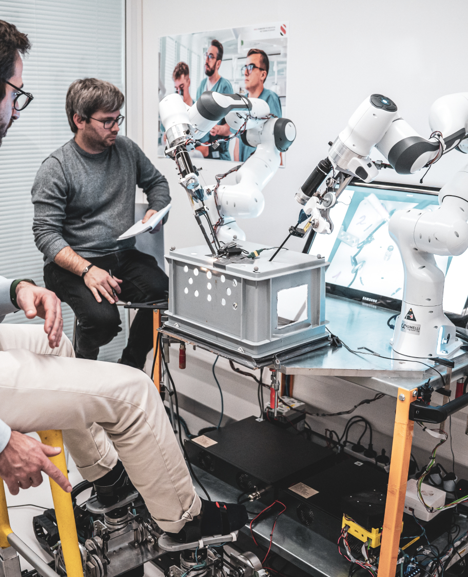
Surgical Robotics, A curated analysis
Our intern Bryan Kheirallah recently graduated Engineer, after successfully presented his master’s thesis at the EPFL. In collaboration with the EPFL and the SFITS, he built a free and resourceful website on surgical robots.
Interactive, rich, dynamic entry-point to all useful information, this platform lets you review and search complete data sheets about surgical robots: description, specialties of use, characteristics, analytical data, feedback from surgeons. Every robot listed on this platform is marketed, CE marked and/or FDA approved.
His master’s thesis was based on the “State of the Art in Robotic Surgery”, an inventory put together in 2021 and revised in 2023 by the Biomedical and Equipment Department of the HUG.
In addition, Bryan deeply studied all existing literature and analyzed surgeons’ feedback on their robotic experiences. He then established a white paper and a checklist of risks for hospital professionals when acquiring a surgical robot.
We take this opportunity to congratulate Bryan for his hard work and his diploma.
The avian model: a novel and cost‑effective animal tissue model for training in neonatal laparoscopic surgery
A prospective observational study, ran by Dr. Peter Zimmermann, Mrs. Ashley Xavérine Wiseman, Dr. Oliver Sanchez, Dr. Amulya K. Saxena, Dr. Enrico Brönniman, was performed during two Minimally Access Surgery Skill Labs in June 2018 and April 2019. It aimed to design and validate a new and cost-effective animal tissue model for training neonatal minimal access surgery (MAS) skills. Selected laparoscopic exercises were performed on organic pieces using 3mm MAS instruments (adhesiolysis, cholecystectomy and intestinal anastomosis). Twenty-seven course participants were recruited (18 females: 9 males) all with different levels of experience. At the end of the tests, the novel avian tissue model for neonatal MAS training could be validated with success.