October 30, 2025 - News
Les news “SFITS Insight”
Les news “SFITS Insight”
Une fois par mois, nous publions sur nos réseaux sociaux (LinkedIn & Instagram) un texte instructeur et/ou informatif écrit par un.e chirurgien.ne qui collabore avec la SFITS. Nous souhaitons profiter du savoir de nos intervenant.e.s pour partager à notre communauté une anecdote ou technique chirurgicale. Étant éparpillées parmi toutes nos publications, nous vous les avons réuni ci-dessous.
Vous avez participé ou donné des cours à la SFITS et vous désirez nous faire parvenir quelques lignes ?
Historique des dernières news
l’Audiologie: une spécialité qui écoute, équilibre… et innove ! (Octobre 2025)
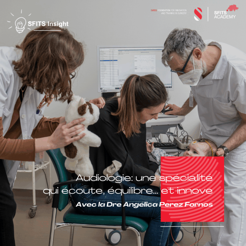
La Dre Angélica Perez Fornos, Ingénieure responsable du Centre Universitaire Romand d’Implants Cochléaires et du laboratoire d’audiologie et vestibulométrie des Hôpitaux Universitaires de Genève (HUG) nous présente “l’Audiologie: une spécialité qui écoute, équilibre… et innove !”
L’audiologie moderne évolue rapidement :
- Nouveaux profils de patients (surdités unilatérales, exposition excessive au bruit, enfants avec troubles vestibulaires…)
- Nouvelles techniques comme l’audiométrie haute fréquence, localisation sonore et les tests dans le bruit, qui permettent de mieux refléter la réalité des symptômes et de diagnostiquer des entités avant méconnues comme la perte auditive cachée.
- Les troubles vestibulaires, longtemps sous-estimés, gagnent en visibilité. Vertiges, instabilité, troubles visuels et cognitifs : leurs symptômes altèrent profondément la qualité de vie. Grâce à des outils diagnostics plus précis (vidéo Head Impulse Test, VEMP, etc.), nous identifions mieux ces pathologies et les intégrons dans nos réflexions cliniques.
Les atteintes de l’oreille interne, qu’elles soient auditives ET vestibulaires, nécessitent une approche globale :
- Tests génétiques pour mieux comprendre les causes et personnaliser les traitements.
- Dépistage précoce y compris vestibulaire (crucial chez les enfants atteints de surdité).
- Formation spécialisée des audiologistes, essentielle pour une prise en charge de qualité.
- Collaboration multidisciplinaire (ORL, neurologues, logopédistes, physiothérapeutes, etc.) au cœur du parcours patient.
Et demain ? Thérapie génique, implants vestibulaires, IA embarquée… L’implant cochléaire, pionnier des neuroprothèses, a ouvert la voie à une révolution technologique qui dépasse l’audition.
En résumé, l’audiologie d’aujourd’hui est une spécialité au croisement de la médecine, de la technologie et de l’humain.
the rise of POCUS (Point-of-Care Ultrasound) (Septembre 2025)
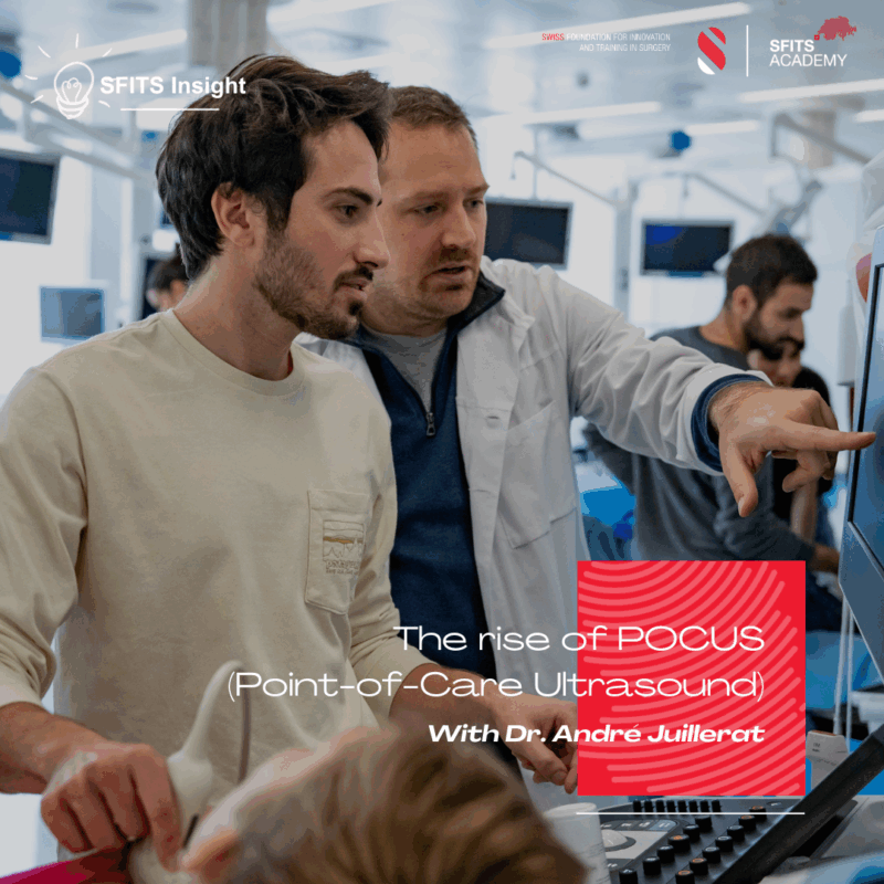
Dr. André Juillerat, chief resident in Internal Medicine at the Geneva University Hospitals (HUG), shares his perspective on the rise of POCUS (Point-of-Care Ultrasound), an ultrasound performed directly at the patient’s bedside using portable – or even hand-held – devices.
#POCUS is transforming clinical practice by providing immediate and targeted answers to binary clinical questions. Long considered a tool limited to certain specialties, ultrasound mastery is now becoming an essential skill for many hospital-based physicians… and why not for surgeons?
Numerous studies have shown that POCUS can be effectively taught to surgeons, improving diagnostic accuracy, reducing delays, and even enhancing patient outcomes. Building on this evidence, an introductory POCUS workshop for surgeons was launched in January 2023 at the SFITS, in its Cours de base de la SFITS Academy. It combines e-learning with hands-on practice on healthy volunteers, supported by simulation to illustrate pathological cases. The workshop was very well received, and participants expressed strong interest in further training.
For surgeons, POCUS could make a real difference in cases such as suspected cholecystitis, pleural effusion, or pneumothorax, to name just a few. In peripheral hospitals, where 24/7 access to radiology is not always guaranteed, equipping surgeons with ultrasound skills may prove particularly valuable.
Recently, HUG established the Unit of Ultrasound in Medicine (U2M), led by Dr. Olivier Grosgurin, dedicated to research, teaching, and educational innovation in POCUS. Among its flagship projects, U2M has developed 3D-printed heart models, in collaboration with the Anatomy Unit of the Faculty of Medicine, to support focused cardiac ultrasound courses. These models help learners grasp the complex visuospatial orientation of echocardiographic views and accelerate their practical mastery.
POCUS exemplifies how an accessible technology can strengthen clinical reasoning, enhance patient safety, and transform medical education.
Breathing Without Breathing: High-Flow Oxygen and the THRIVE Technique (Juin 2025)
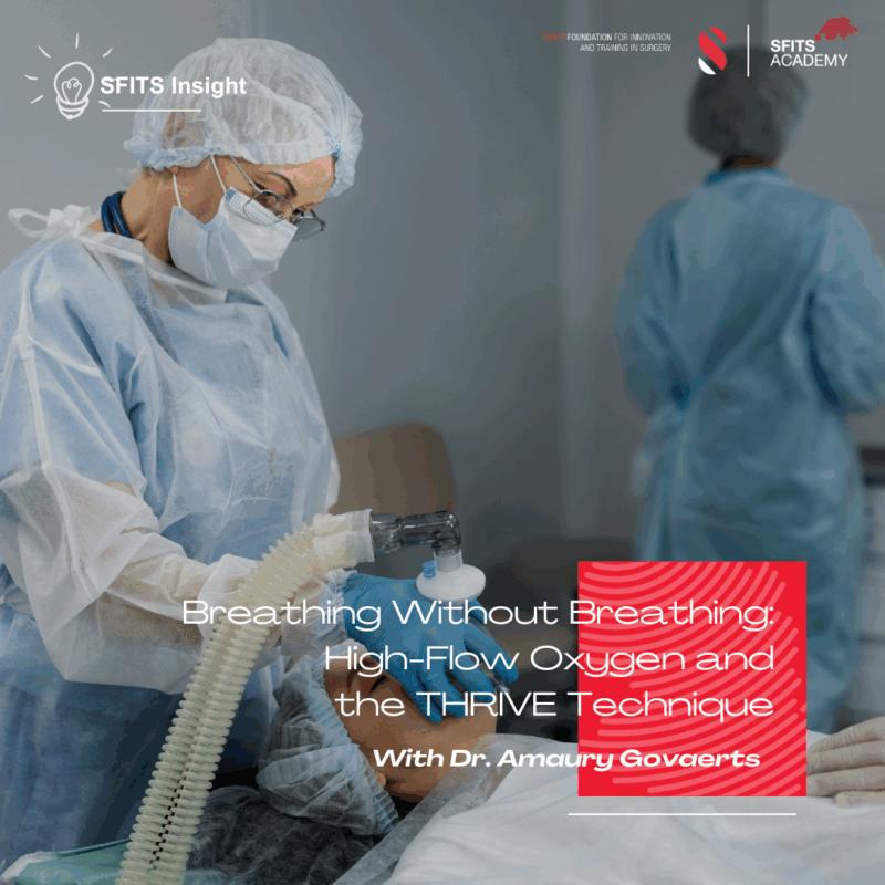
Dr. Amaury Govaerts, Physician assistant at the Geneva University Hospitals would like to tell us more about Breathing Without Breathing: High-Flow Oxygen and the THRIVE Technique Spotlight on Anesthesia Practice.
In certain surgeries — especially hashtagENT procedures involving the airway or face — placing a breathing tube (intubation) can obstruct the surgical field. A remarkable alternative, called apneic oxygenation, allows oxygen to continue flowing into the lungs even when the patient is fully asleep and isn’t actively breathing— either spontaneously or through a ventilator. Delivered through a soft nasal interface at high flow rates (up to 60 L/min), this counterintuitive concept works because oxygen continues to diffuse passively into the bloodstream as long as it is supplied under pressure, even without visible chest movement. However, carbon dioxide (CO2) is not removed during this process, so it gradually accumulates — limiting how long apneic oxygenation can be safely maintained.
A major advance in this area is the hashtagTHRIVE technique (Transnasal Humidified Rapid-Insufflation Ventilatory Exchange), developed by Dr. Anil Patel in 2015. THRIVE uses warm, humidified, high-flow oxygen to extend the apneic window — sometimes up to 30 minutes or more in carefully selected patients. This has transformed how anesthesiologists manage difficult airways and short hashtagENT procedures, offering a clear, device-free field for the surgeon and giving teams more time in high-risk airway situations.
At the University Hospitals of Geneva, we use THRIVE routinely for ENT procedures requiring a device-free airway, providing a high level of safety for patients and excellent operating conditions for surgeons. This technique illustrates how innovation, physiology, and teamwork converge in modern anesthesia to push the boundaries of what’s possible in surgical care.
Ref: Patel A, Nouraei SAR. Transnasal Humidified Rapid-Insufflation Ventilatory Exchange (THRIVE): A physiologically based apnoeic oxygenation technique. Anaesthesia. 2015;70(3):323–329. doi:10.1111/anae.12923
Submucous Cleft Palate (Avril 2025)
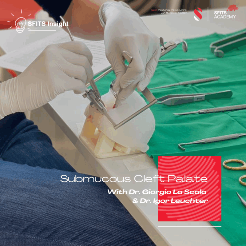
Dr. Giorgio La Scala, Associate Physician in the Pediatric surgery unit and Dr. Igor Leuchter, Associate Physician in the Departement of Otorhinolaryngology, both working at the Geneva University Hospitals, are giving us interesting details about submucous cleft palate.
Some patients may have speech that sounds unusually nasal, which is often linked to a cleft palate—even when the palate appears normal. In some cases, the voice was normal until after an adenoidectomy. Other patients might only notice nasal air escape when blowing forcefully, such as when playing a wind instrument.
Although muscle weakness from certain neurological conditions can cause similar issues, the underlying problem can be an anatomical condition called occult submucous cleft palate, caused by malposition of the muscles in the palate. This condition can only be diagnosed with a nasofibroscopy. If the symptoms significantly affect quality of life, treatment options may include speech therapy or surgery. Surgical approaches vary depending on the severity and may involve fat grafting (lipofilling) to the back of the throat (posterior pharyngeal wall), reconstruction of the palatal muscles sling (palatoplasty), or even lengthening of the palate.
At the HUG, the ENT Phoniatry Clinic and the Pediatric Plastic Surgery Clinic work together to offer specialized diagnosis and treatment for both children and adults affected by this condition.
L'utilisation du Fluoptics lors des mastectomies (Mars 2025)
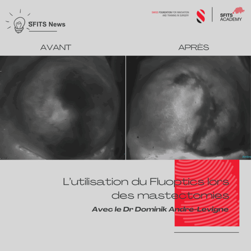
Le Dr Dominik André-Lévigne, chef de clinique dans le Service de Chirurgie plastique, reconstructive et esthétique des Hôpitaux Universitaires de Genève nous parle de l’utilisation du Fluoptics lors des mastectomies à préservation cutanée ou de mastectomies à préservation du mamelon suivi d’une reconstruction immédiate par prothèse.
Cette technique permet de visualiser la vascularisation relative de la peau pendant l’intervention chirurgicale grâce à l’injection de vert d’indocyanine (fluorescence). Elle permet ainsi de prévenir l’exposition des implants lors d’une reconstruction mammaire immédiate, en identifiant précocement les zones à risque de nécrose cutanée.
On peut distinguer les zones bien vascularisées, celles présentant un retard de vascularisation, et celles totalement dépourvues de vascularisation. Le Dr André-Lévigne et son équipe mènent actuellement une étude sur l’utilité du caisson hyperbare pour sauver les zones montrant un retard de vascularisation à la fluorescence.
Cette visualisation peropératoire de la vascularisation cutanée permet une exérèse précise des tissus non vascularisés, ce qui optimise les résultats esthétiques de la reconstruction immédiate et réduit le risque de reprise chirurgicale liée à des souffrances cutanées.
A new hope for patient with symptomatic levator ani avulsion (Février 2025)
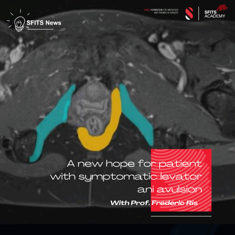
Prof. Frédéric Ris, Associate Physician, at the Geneva University Hospitals (HUG) tells us more about patient with symptomatic levator ani avulsion.
Levator Ani Avulsion is a common pathology, occuring after delivery in 7% of the women population and up to 50% of forceps deliveries. This can lead to a wide variety of symptoms, with anal dyschesia or incontinence, urine incontinence, colpophonia (air from the vagina), pain during sexual intercourse, or loss/decrease of orgasm. It is often seen with a feeling of musculoskeletal unbalance and widening of the vagina. It is believed that this condition cannot be repaired by a classical urogynecological vision.
We developped in our Proctology Unit of the HUG a new operation to solve this issue in symptomatic patients with improvement or complete resolution of the symptoms in most of the patients.
This intervention aims at restoring the anatomy of the pelvic floor, and the balance without foreign body, just simple stiches. Complication rate is low and patient can go home after a short hospital stay.
As a leading unit for this surgery, we tend to teach it in Switzerland or accross countries in Europe or to welcome patients from all over the world.
Faut-il utiliser des clamps en microchirurgie (Janvier 2025)
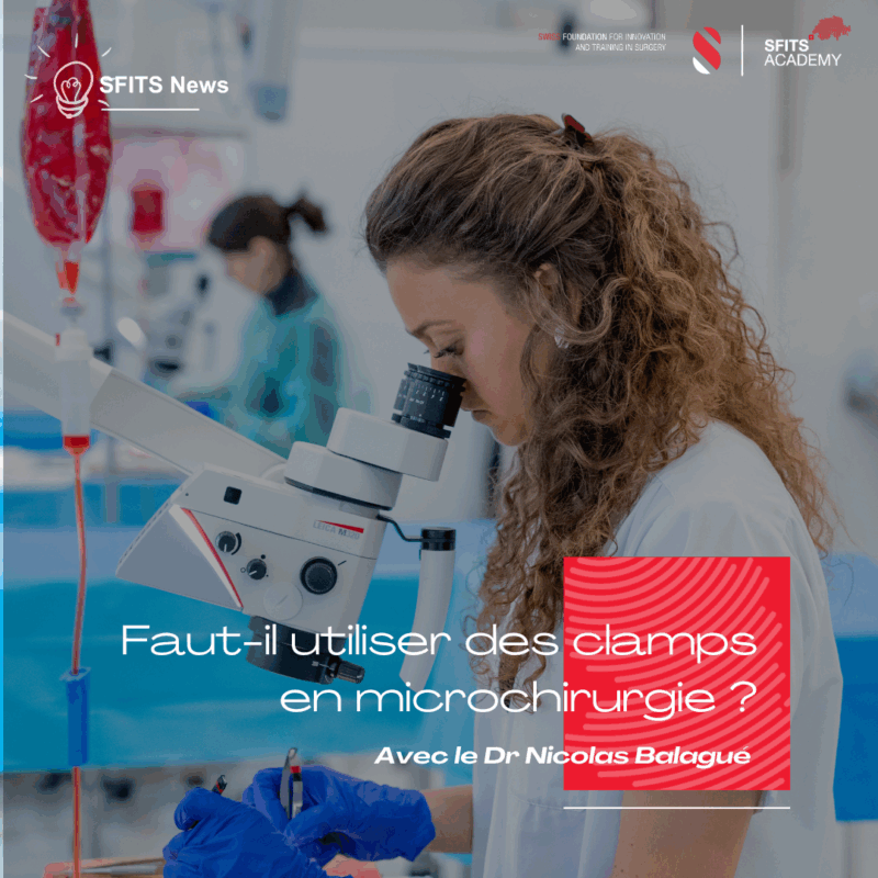
Le Dr Nicolas Balagué, médecin chef de service en Chirurgie plastique à l’Hôpital de Sierre, nous explique.
❓ Faut-il utiliser des clamps en microchirurgie ?
La microchirurgie associe la chirurgie vasculaire à l’utilisation d’un microscope. Le microscope facilite la suture de vaisseaux dont le diamètre est inférieur à 3 mm (et pouvant aller jusqu’à 0.3 mm). Pour les vaisseaux de plus gros calibre, l’utilisation de loupes est souvent suffisante.
En 1966, Yoshikazu Ikuta, un chirurgien japonais, développait un clamp vasculaire en métal qui permettait de faciliter la disposition des vaisseaux pour pratiquer la suture. Depuis, bien d’autres clamps ont été développés, certains ayant une pression variable selon le calibre du vaisseau et sa nature (veine ou artère). Toutefois, l’utilisation de ces clamps varie d’une spécialité à l’autre et d’un hôpital à l’autre.
Ainsi, la plupart des chirurgiens en plastique utilisent des clamps pour effectuer les anastomoses des lambeaux, tandis que les chirurgiens de la main (qui travaillent sur une surface souvent plane avec un espace anatomique réduit) ont l’habitude d’effectuer les sutures vasculaires sans clamps.
L’utilisation du clamp influence également la technique de la suture puisque le retournement du vaisseau imposé par le clamp nécessite une suture en 2 temps (selon Ikuta), tandis que l’impossibilité de retourner un vaisseau (sur un doigt par exemple) nécessite d’utiliser une autre technique de suture de la profondeur à la superficie (selon Comptet).